Heart Imaging: What It Is, How It Works, and What It Reveals
When your doctor suspects something’s off with your heart, they don’t guess—they look. Heart imaging, a group of noninvasive techniques that create detailed pictures of the heart’s structure and function. Also known as cardiac imaging, it’s how doctors spot blocked arteries, weak muscles, faulty valves, and abnormal rhythms before they cause a crisis. This isn’t science fiction—it’s routine care for millions, and it’s why more people survive heart problems today than ever before.
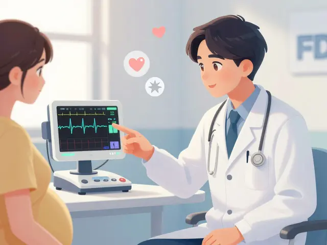
There are several ways to image the heart, each with a different strength. An echocardiogram, a type of ultrasound that uses sound waves to show the heart beating in real time is often the first step. It’s safe, quick, and shows if the heart is pumping well or if valves are leaking. If something’s still unclear, a CT angiogram, a scan that uses X-rays and contrast dye to map blood flow through coronary arteries can reveal blockages you can’t see any other way. For detailed tissue analysis—like scar tissue from a past heart attack—MRI heart, a powerful magnetic scan that gives ultra-clear images of heart muscle and blood flow is the gold standard. These aren’t interchangeable tools; each answers a different question.
Heart imaging doesn’t just diagnose—it guides treatment. A low ejection fraction seen on an echocardiogram might mean you need a pacemaker. A blockage caught on a CT scan could lead to a stent. An MRI showing scar tissue might change your medication plan. These tests also help track progress. Did your heart function improve after starting a new drug? Was the surgery successful? Imaging gives the answer. And because many of these scans don’t require cuts or long recovery, they’re used again and again over time.
What you won’t find in most doctor’s offices is a one-size-fits-all approach. The choice of test depends on your symptoms, risk factors, and what the doctor suspects. Someone with chest pain and high cholesterol gets a different scan than someone with irregular heartbeats or shortness of breath after walking. That’s why understanding what each test does matters—it helps you ask the right questions and know what to expect.
Below, you’ll find real-world guides on how heart imaging connects to medications, risk scores, and lifestyle decisions. From how ASCVD scores influence whether you need a stress test, to how drug interactions affect heart function, these posts show you how imaging fits into the bigger picture of heart health. No fluff. Just what you need to understand your results, your options, and your next steps.

Cardiac MRI vs Echocardiography: Which Heart Scan Gives You the Real Picture?
Cardiac MRI and echocardiography are both vital for heart imaging, but they serve different roles. Echocardiography is fast and widely used for initial checks, while cardiac MRI offers unmatched detail for tissue and function analysis-especially when echo results are unclear.